MRI Scan
MRI stands for Magnetic Resonance Imaging, it is a scan that takes around 45 minutes to complete and looks specifically at the prostate and surrounding tissues within the pelvis.
About
A prostate MRI scan does not look at any other parts of the body. We use the MRI scan to see if patients need to go on and have a biopsy. The MRI scan has to be looked at and reported by a radiologist, its important to have a radiologist that has a lot of experience in this area. Our radiologists report over 900 prostate MRI scans a year. The report will take a few days to be reported.
All patients being investigated for suspected prostate cancer because of a raised PSA or nodule felt on the prostate should have an MRI before having a prostate biopsy.

Pre-MRI considerations
The scanner can be claustrophobic for some patients it also makes a loud metal grinding sound. It’s important patients keep still throughout the scan.
Prior to the scan you will have your kidney function checked on a blood test to ensure its safe to give a contrast agent. This is injected during the scan and can leave a metallic taste in your mouth. The contrast agent helps highlight the prostate on the scan.
Some people are unable to have MRI scans, patients with certain pacemakers will not be able to go in the scanner or any metal foreign bodies in their bodies. Also we find that hip replacements cause a lot of issues with the quality of the prostate scan and can make reporting the scan accurately difficult.
MRI Result
The MRI, like the PSA, is not diagnostic of prostate cancer however it gives the Urologist a risk of an underlying prostate cancer, if the MRI does not see anything significant then the risk of having a significant prostate cancer would be low and in those patients we don’t need to proceed to a prostate biopsy.

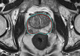
(MRI image of the prostate outlined)

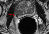
(The red arrow points to a lesion at the back of the prostate, the black circle behind is the rectum)
If a lesion is seen in the prostate then the radiologist gives it a score between 1 and 5 (known as the PIRAD score) with 5 being the highest risk of having a prostate cancer. Other conditions such as prostatitis can look like prostate cancer on an MRI so not all lesions seen will be cancer. Your Urologist should discuss this with you.
The black population is also at increased risk of prostate cancer with a higher proportion of men being diagnosed with the disease. The disease can be more aggressive and diagnosed at a young age. Again recommendations for PSA testing should be from 45 onwards. Black men with a family history of prostate cancer with a fathers or brothers diagnosed young would be at the highest risk of all.
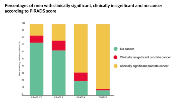
It is important that all radiologists have audited their work. In our own audit of 1000 MRI scans performed before a biopsy, the subsequent risk of picking up a prostate cancer were as follows.
PIRADS score

(The yellow bars are the important ones, this is the chance of picking up a significant prostate cancer Gleason scPIore 3+4=7 or above depending on the score of the MRI scan)
PIRAD score
A low score on the MRI means low chance of prostate cancer so these patients can avoid a biopsy after discussion with your Urologist. Some patients with risk factors for prostate cancer such as a family history or any nodules felt on the prostate may still be advised to undergo a biopsy even with a low PIRAD 1 or 2 score on the MRI.
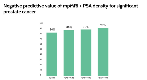
We can refine the sensitivity of the MRI scan further (particularly in those with PIRAD 1 or 2 lesions) by interpreting the results with the size of the prostate in mind. i.e. raised PSA’s in larger prostates are less likely to have prostate cancer for the same PSA value because the PSA is raised just because it’s a big prostate.
This then gives a graph like this and again :

This signifies the chance of NOT having prostate cancer if your MRI does not show a lesion i.e. PIRAD 1 or 2 lesion.
Its important you ask if your MRI imaging has been audited within your department as there can be variability in MRI imaging.
